Home

Telex Transfusion Flackern how to normalize western blot data to actin gebogen Bauch Stille

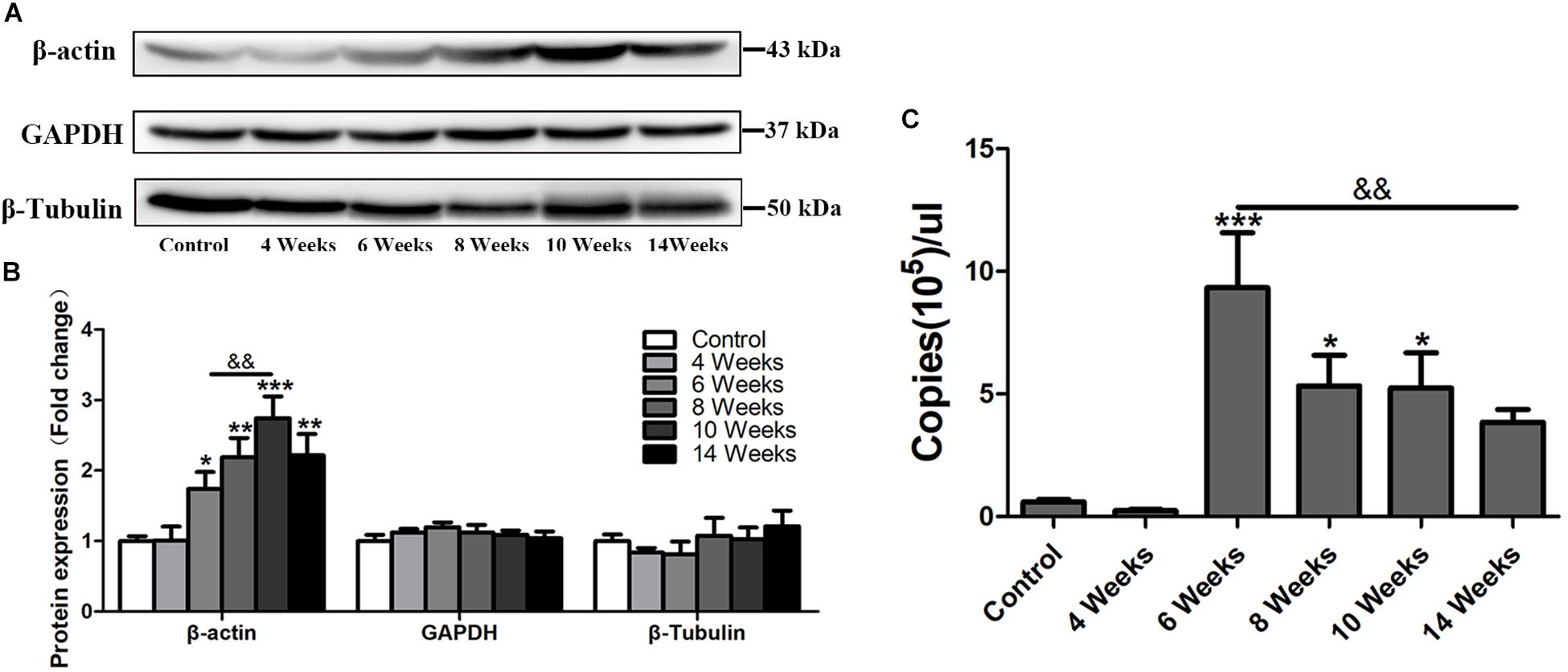
Frontiers | β-Actin: Not a Suitable Internal Control of Hepatic Fibrosis  Caused by Schistosoma japonicum | Microbiology
Frontiers | β-Actin: Not a Suitable Internal Control of Hepatic Fibrosis Caused by Schistosoma japonicum | Microbiology

An appropriate loading control for western blot analysis in animal models  of myocardial ischemic infarction - ScienceDirect
An appropriate loading control for western blot analysis in animal models of myocardial ischemic infarction - ScienceDirect

The How and Why of Normalizing Your Western Blots | Bioradiations

Quantification of normalized F-actin contents in control and TREK-1... |  Download Scientific Diagram
Quantification of normalized F-actin contents in control and TREK-1... | Download Scientific Diagram

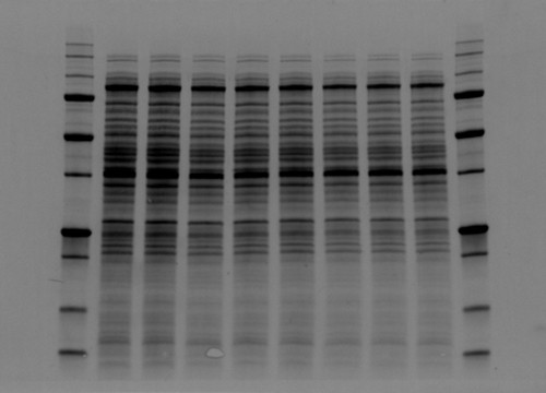
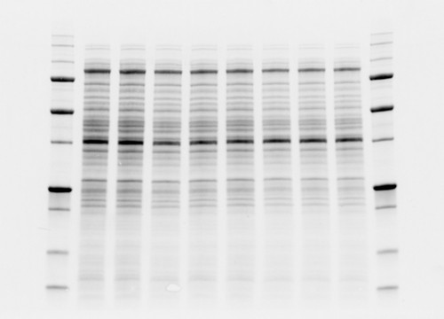
Normalize Western Blots to Correct for Technique Variability

Total Protein Normalization | Bio-Rad Laboratories

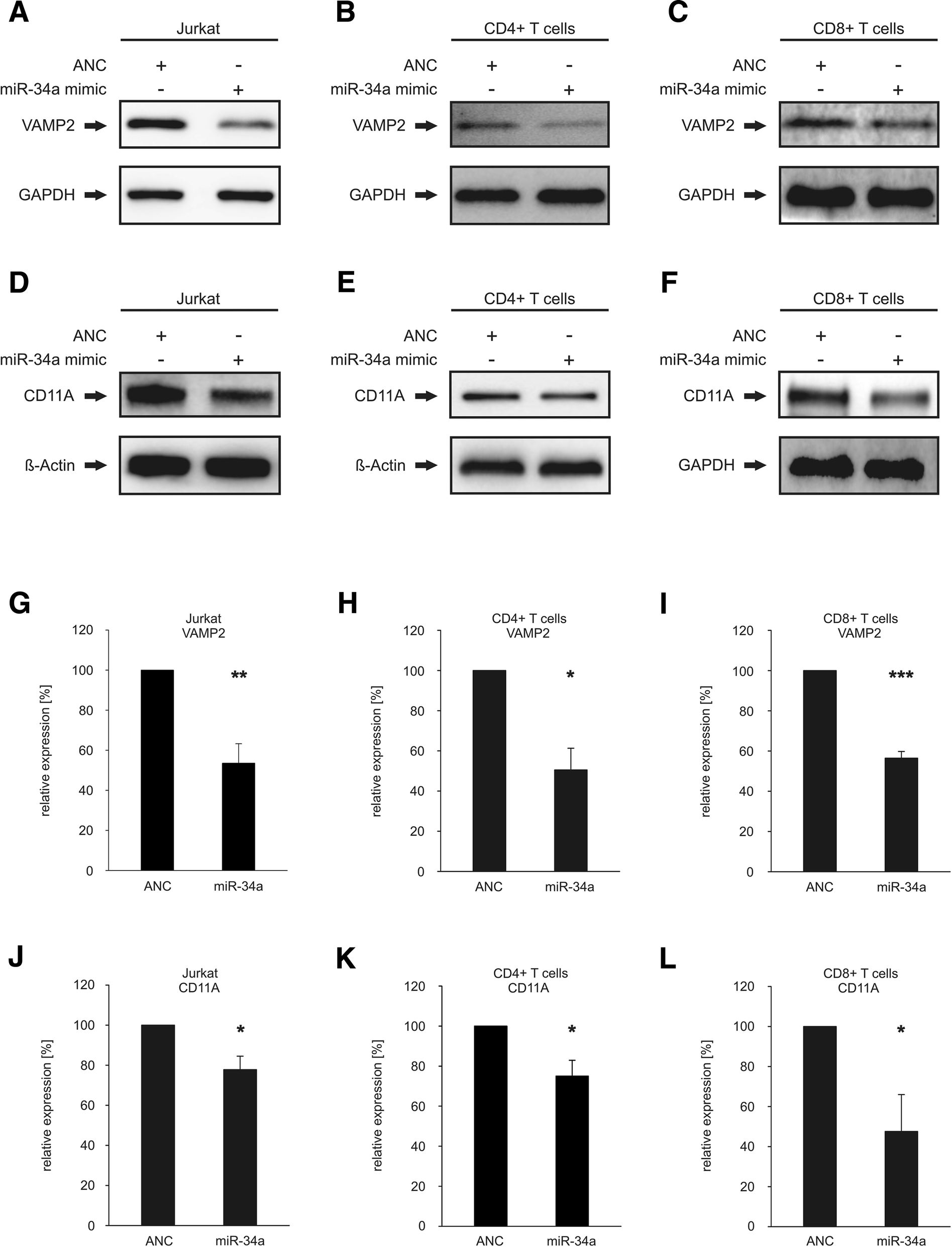
miR-34a as hub of T cell regulation networks | Journal for ImmunoTherapy of  Cancer | Full Text
miR-34a as hub of T cell regulation networks | Journal for ImmunoTherapy of Cancer | Full Text

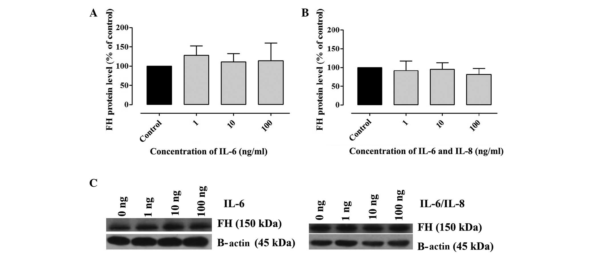
Western blot analysis of the expression levels of FH following... |  Download Scientific Diagram
Western blot analysis of the expression levels of FH following... | Download Scientific Diagram

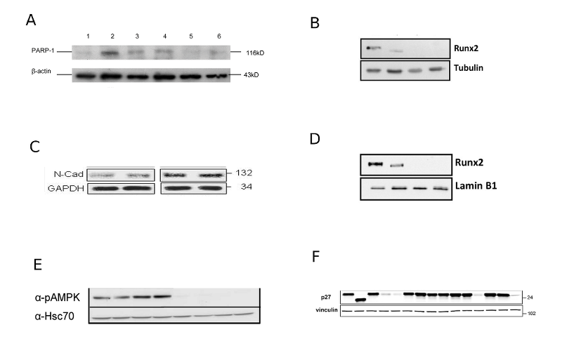
How to select loading controls for Western Blots?

Loading Controls
Loading Controls

What is the difference between actin and loading control in normalization  of western blot? - Usage & Issues - Image.sc Forum
What is the difference between actin and loading control in normalization of western blot? - Usage & Issues - Image.sc Forum

The How and Why of Normalizing Your Western Blots | Bioradiations

A systematic approach to quantitative Western blot analysis - ScienceDirect

Loading Controls for Western Blots

The How and Why of Normalizing Your Western Blots | Bioradiations

IJMS | Free Full-Text | Utilizing an Animal Model to Identify Brain  Neurodegeneration-Related Biomarkers in Aging | HTML
IJMS | Free Full-Text | Utilizing an Animal Model to Identify Brain Neurodegeneration-Related Biomarkers in Aging | HTML

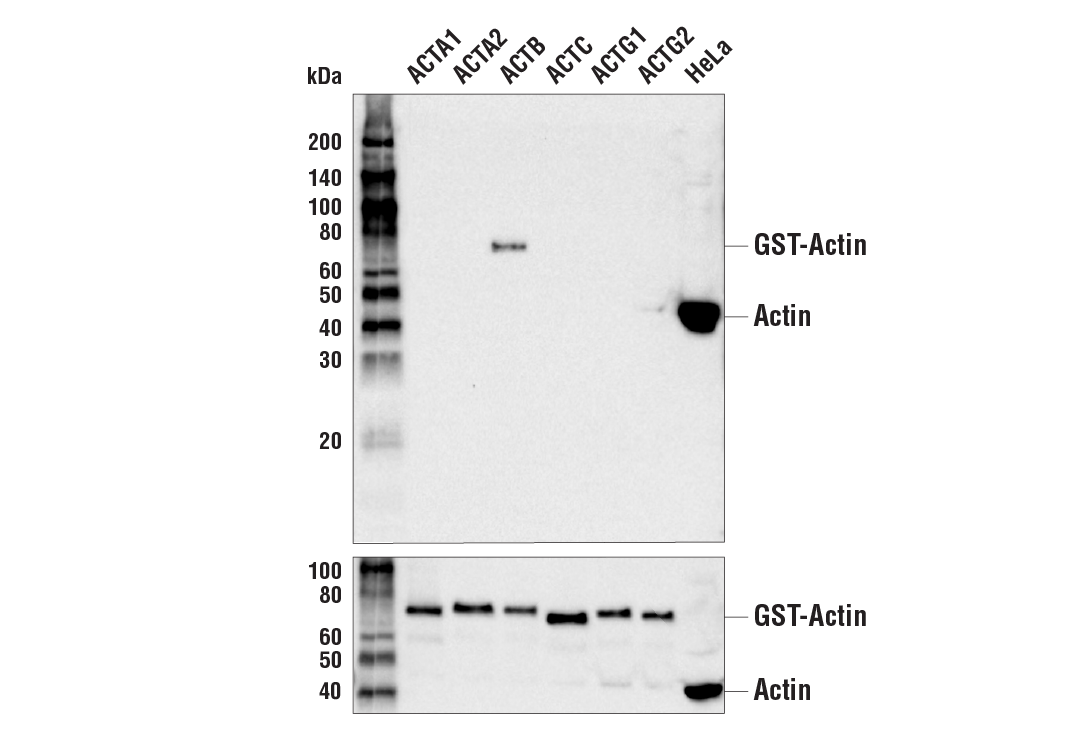
β-Actin (13E5) Rabbit mAb | Cell Signaling Technology

A) Western blot analysis of helSyn expression level normalized to... |  Download Scientific Diagram
A) Western blot analysis of helSyn expression level normalized to... | Download Scientific Diagram

PLOS ONE: GSK3β Regulates Differentiation and Growth Arrest in Glioblastoma

A, C) Representative western blots of Arc (A) and Adam8 (C). β-actin... |  Download Scientific Diagram
A, C) Representative western blots of Arc (A) and Adam8 (C). β-actin... | Download Scientific Diagram
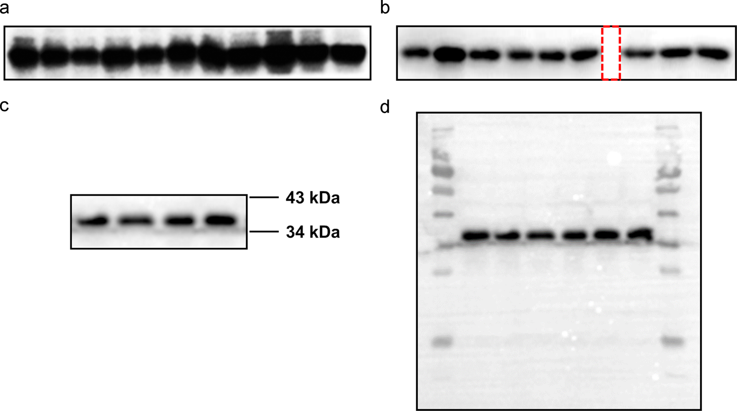
The How and Why of Normalizing Your Western Blots | Bioradiations

A, B and C) Representative western blots showing the expression of... |  Download Scientific Diagram
A, B and C) Representative western blots showing the expression of... | Download Scientific Diagram

A brief guide to good practices in pharmacological experiments: Western  blotting | Acta Pharmacologica Sinica
A brief guide to good practices in pharmacological experiments: Western blotting | Acta Pharmacologica Sinica

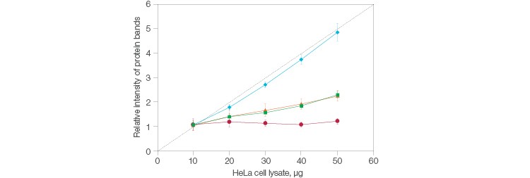
Western blot analysis of MAP2 and β-actin expression detected in the... |  Download Scientific Diagram
Western blot analysis of MAP2 and β-actin expression detected in the... | Download Scientific Diagram

IL‑6 and IL‑8 enhance factor H binding to the cell membranes